
Immediately, chest X-ray images can now be analyzed using artificial intelligence to detect potential lung cancer symptoms in individuals who do not smoke, potentially identifying those at higher risk.
Lung cancer, the primary cause of cancer-related fatalities globally, is attributed to smoking in 85% of cases. However, factors such as exposure to “second-hand smoke,” radon, asbestos, air pollution, and a family history of lung cancer can render 15% of the population susceptible to the disease.
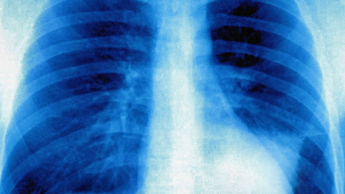
A recent study presented at the annual meeting of the Radiological Society of North America has revealed that machine learning algorithms can be trained to identify signs of lung cancer in X-ray images. A comparison of X-ray images shows a small nodular opacity in the left upper lung zone, detected by AI, contrasting with a solid nodule detected in the upper lobe by a low-dose, non-contrast chest CT scan. The study was credited to RSNA/Anika S. Walia.
This advancement could potentially provide a simple screening method for individuals who have never smoked but may still be at risk due to other contributing factors.
In the United States, authorities recommend lung cancer screening for individuals aged 50 to 80 with a history of smoking at least 20 packs per month. Australia is set to implement similar screening protocols starting in July 2025.
While not currently recommended for non-smokers, the screening process, typically conducted through low-dose CT scans, boasts high sensitivity and specificity in detecting cancer. Anika Walia, a researcher at Boston University School of Medicine, believes that AI could play a crucial role in ensuring these individuals are not overlooked.
Walia led a study that trained a deep learning model, named “CXR-Lung-Rise,” to identify cancer-related patterns in chest X-rays. This approach is more cost-effective than using CT scans.
The model was trained on over 150,000 chest X-rays from more than 40,000 asymptomatic smokers and non-smokers participating in a comprehensive cancer screening program in the US to assess lung cancer risk.
Approximately 28% of the participants were classified as high-risk for developing lung cancer, with 2.9% subsequently receiving a diagnosis of the disease.
Dr. Michael Lu, AI director at the Cardiovascular Imaging Research Center at Massachusetts General Hospital, believes this discovery paves the way for novel screening methods as smoking rates decline.
Lu stated, “By utilizing existing chest X-rays in electronic health records, this AI tool enables proactive screening for never-smokers at elevated risk of lung cancer.”
Given the decreasing rates of cigarette smoking, strategies for early lung cancer detection in non-smokers are becoming increasingly vital.