Time-constrained medical professionals may benefit from a novel artificial intelligence tool designed to enhance the analysis of illnesses and the interpretation of clinical images with exceptional clarity.
The iStar (Inferring Super-Resolution Tissue Architecture) device, developed by researchers at the University of Pennsylvania’s Perelman School of Medicine, aims to assist clinicians in identifying and treating cancers that might otherwise remain undetected.
By utilizing advanced imaging techniques, doctors and researchers can now visualize tumor cells that were previously elusive, gaining comprehensive insights into adult cell structures and the intricate workings of genetic processes. This innovative tool offers rapid annotation of micro images and facilitates the evaluation of surgical margins in cancer procedures, paving the way for precise molecular disease diagnostics.
A study led by Daiwei “David” Zhang, Ph.D., and Mingyao Li, Ph.D., a specialist in Biostatistics and Digital Pathology, was recently published in Nature Biology, highlighting the capabilities of iStar.
Li emphasizes that iStar’s ability to swiftly detect “tertiary lymphatic structures,” crucial immune formations linked to patient survival rates and response to immunotherapy, can aid in personalized treatment decisions for cancer patients.
The development of iStar involved the integration of geographic transcriptomics, a cutting-edge technique for mapping protein activities in tissue regions, and the adaptation of the Hierarchical Vision Transformer, a machine learning tool, by Li and her team.
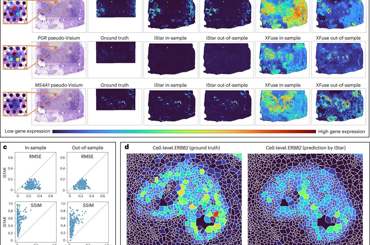
Through a meticulous process of image dissection and analysis, iStar leverages AI algorithms to decipher gene activities at a near-single-cell resolution, providing a comprehensive understanding of tissue structures and cellular patterns akin to a pathologist’s examination of a muscle sample.
In experimental trials involving various cancer cell types and healthy tissues, iStar demonstrated remarkable proficiency in identifying elusive tumor cells, offering promise for improved cancer detection and diagnosis.
Notably, iStar outperformed other AI tools in terms of speed, completing evaluations in a fraction of the time while maintaining accuracy and efficiency. This accelerated pace is crucial for conducting extensive medical research and analyzing complex 3D tissue data and biobank repositories.
Li and her team are committed to further enhancing iStar’s capabilities to enable in-depth exploration of cellular microenvironments, ultimately enhancing diagnostic accuracy and therapeutic outcomes in medical practice.


