by Roberto Molar Candanosa, Johns Hopkins University
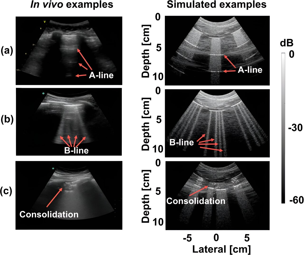
 In both live and simulated scenarios, examples of (a) A-line, (b) B-line, and © consolidation features are showcased. Credit: Communications Medicine (2024). DOI: 10.1038/s43856-024-00463-5
Recent research suggests that artificial intelligence has the capability to identify COVID-19 in lung ultrasound images akin to how facial recognition software distinguishes faces in a group.
These findings enhance AI-powered clinical diagnostics, empowering healthcare professionals to better detect COVID-19 and other respiratory ailments using algorithms that analyze ultrasound images to detect disease indicators.
The study, recently published in Communications Medicine, marks the culmination of a project initiated early in the pandemic to address the urgent need for rapid patient assessment in overcrowded emergency rooms.
Muyinatu Bell, the lead author and John C. Malone Associate Professor of Electrical and Computer Engineering, Biomedical Engineering, and Computer Science at Johns Hopkins University, stated, “We devised this automated monitoring tool to assist healthcare providers in high-pressure environments with a large volume of patients requiring swift and accurate diagnoses. Additionally, we envision the potential for mobile devices that patients can utilize at home to monitor the progression of COVID-19.”
The tool also holds promise for monitoring conditions like congestive heart failure, which can lead to fluid accumulation in the lungs, as mentioned by co-author Tiffany Fong, an assistant professor of emergency medicine at Johns Hopkins Medicine.
Fong emphasized that leveraging AI tools represents the “next major frontier” in point-of-care applications. For instance, wearable ultrasound patches could monitor fluid retention and alert individuals to seek medical attention or adjust their medication.
The AI system focuses on identifying B-lines in lung ultrasound images, which are bright, vertical patterns signaling inflammation in patients with pulmonary complications. By integrating computer-generated images with actual patient ultrasounds, including those from Johns Hopkins, the system aims to enhance diagnostic accuracy.
Bell explained, “We needed to accurately model ultrasound physics and acoustic wave propagation to generate realistic simulated images. Subsequently, we trained our computer models to interpret real lung scans of patients with compromised lung function using this simulated data.”
At the onset of the pandemic, the lack of patient data and understanding of the disease’s physiological manifestations posed challenges for utilizing AI to interpret COVID-19 indicators in lung ultrasound images, Bell noted.
To address this, the team developed software capable of learning from a combination of real and simulated data to detect abnormalities in ultrasound scans indicative of COVID-19 infection. This software employs a deep neural network, a form of AI that mimics the information processing capabilities of interconnected neurons in the human brain, enabling it to recognize patterns and interpret complex data.
First author Lingyi Zhao, who created the software during their postdoctoral tenure in Bell’s lab and is currently affiliated with Novateur Research Solutions, remarked, “During the initial stages of the pandemic, the scarcity of ultrasound images from COVID-19 patients hindered the development and testing of our algorithms, leading to suboptimal performance of our deep neural networks. However, we are now demonstrating that computer-generated datasets can effectively identify COVID-19 features with high accuracy.”
More information: Lingyi Zhao et al, Detection of COVID-19 features in lung ultrasound images using deep neural networks, Communications Medicine (2024). DOI: 10.1038/s43856-024-00463-5
Provided by Johns Hopkins University
Citation: AI can now detect COVID-19 in lung ultrasound images (2024, March 20) retrieved 20 March 2024 from https://medicalxpress.com/news/2024-03-ai-covid-lung-ultrasound-images.html
Copyright © 2024 Johns Hopkins University. Reproduction prohibited without permission, except for fair use for private study or research purposes. Information provided for informational purposes only.


